Obstructive azoospermia is a common cause of male infertility where sperm production is normal, but a blockage prevents sperm from appearing in the semen. This blog explores how radiological techniques—such as scrotal ultrasound, transrectal ultrasound (TRUS), and testicular biopsy imaging—play a vital role in diagnosing this condition. Learn what to expect during these procedures and how expert evaluation can guide effective treatment options.
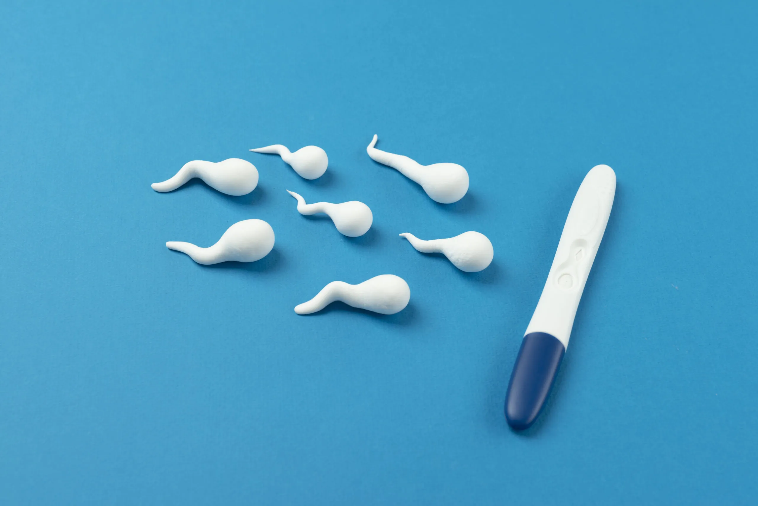
Obstructive azoospermia is a condition where sperm production in the testes is normal, but sperm is unable to reach the semen due to a physical blockage somewhere along the male reproductive tract. While it accounts for up to 40% of cases of azoospermia, this condition often goes unnoticed until a couple struggles to conceive and undergoes fertility testing.
Diagnosing this condition accurately is crucial, as the treatment path for obstructive azoospermia is vastly different from non-obstructive causes. This is where radiological techniques play a vital role. Imaging helps pinpoint the exact location of a blockage, assess the male reproductive anatomy, and provide essential information to guide treatment decisions.
This blog explores the role of radiology in diagnosing obstructive azoospermia and outlines what to expect during these imaging procedures.
Understanding Obstructive Azoospermia
Azoospermia refers to the complete absence of sperm in a man’s semen. It can be broadly divided into two types:
- Non-obstructive azoospermia – when the testicles fail to produce sperm.
- Obstructive azoospermia – when sperm is produced but cannot be transported due to a blockage.
Men with obstructive azoospermia typically have normal hormone levels, and their testicular function is intact. The obstruction can occur in the epididymis, vas deferens, or ejaculatory ducts—anywhere along the pathway where sperm travels from the testicles to the urethra.
Because there are no obvious symptoms, many men discover this condition only through tests done for male fertility evaluation. Radiological diagnosis for male infertility becomes essential in determining the underlying cause of the blockage.
The Role of Radiology in Diagnosing Azoospermia
When a semen analysis shows no sperm, the next step is to find out whether the cause is obstructive or non-obstructive. Radiology offers non-invasive or minimally invasive imaging tests that visualize the male reproductive anatomy, allowing fertility specialists to identify structural abnormalities or blockages.
Let’s take a closer look at the major imaging modalities used in the diagnosis of obstructive azoospermia.
1. Scrotal Ultrasound for Azoospermia
A scrotal ultrasound is often the first imaging test performed when evaluating male infertility. It uses high-frequency sound waves to produce images of the:
- Testicles
- Epididymis
- Vas deferens (if visible)
- Surrounding tissue and blood flow
This test helps identify:
- Abnormalities in sperm production
- Testicular masses or tumors
- Cysts or fluid accumulation (e.g., spermatoceles, hydroceles)
- Signs of inflammation or infection
- Indirect evidence of vas deferens blockage
Because it is quick, non-invasive, and painless, scrotal ultrasound for azoospermia is considered a foundational diagnostic tool in male fertility evaluation.
2. Transrectal Ultrasound (TRUS) for Male Infertility
TRUS, or transrectal ultrasound, offers detailed imaging of the prostate gland, seminal vesicles, and ejaculatory ducts—structures that lie deeper in the pelvic area and cannot be assessed by scrotal ultrasound alone.
TRUS is especially useful when:
- Semen volume is low
- The patient has undergone vasectomy reversal
- There’s suspicion of ejaculatory duct obstruction
- A congenital absence or anomaly in the seminal vesicles is suspected
This test involves inserting a small ultrasound probe into the rectum to capture high-resolution images of the male reproductive tract. Though mildly uncomfortable, TRUS is a vital imaging test for uncovering causes of infertility in men related to deep-seated anatomical issues.
It may reveal:
- Midline prostatic cysts
- Seminal vesicle dilation
- Calcifications or strictures in the ejaculatory ducts
Absence of seminal vesicles (associated with congenital vas deferens abnormalities)
3. MRI and Specialized Imaging (In Select Cases)
While not routinely used in all cases, MRI (Magnetic Resonance Imaging) may be employed when ultrasound findings are inconclusive or when high-resolution soft tissue detail is needed. MRI can detect abnormalities in the pelvis, prostate, or seminal vesicles with greater clarity.
However, due to cost and limited availability, it is usually reserved for more complex diagnostic scenarios.
4. Testicular Biopsy Imaging Guidance
When imaging and blood tests suggest that sperm production is normal but still absent in the semen, a testicular biopsy may be performed. In some cases, this biopsy is image-guided, especially when locating the most active areas of sperm production within the testes.
The biopsy sample helps confirm whether the azoospermia is indeed obstructive and may also be used to retrieve sperm for use in assisted reproductive techniques such as IVF with ICSI.
What Patients Can Expect During Imaging Procedures
For most men undergoing fertility evaluation, imaging procedures are generally well-tolerated and performed on an outpatient basis. Here’s what to expect:
Scrotal Ultrasound:
- Performed externally on the scrotum
- Gel is applied, and a handheld probe is moved across the skin
- Takes about 15–20 minutes
- Painless and requires no preparation
Transrectal Ultrasound (TRUS):
- Requires the patient to lie on their side
- A lubricated ultrasound probe is gently inserted into the rectum
- Mild pressure or discomfort may be felt
- The procedure lasts around 20–30 minutes
Patients are encouraged to discuss any concerns or discomfort with their fertility specialist before the procedure.
Why Radiology Matters in Male Fertility
Radiological imaging is a cornerstone in modern diagnostics, especially when dealing with complex conditions like obstructive azoospermia. The ability to visualize male reproductive anatomy allows clinicians to:
- Differentiate between obstructive and non-obstructive azoospermia
- Identify the location and cause of a blockage
- Decide if surgical correction or sperm retrieval is the best option
- Reduce the need for exploratory procedures
When combined with semen analysis abnormalities, hormonal tests, and physical examination, imaging provides a complete picture of male reproductive health.
Collaborating With a Reproductive Urologist
A reproductive urologist works alongside fertility specialists to manage male infertility. They interpret radiological findings in the context of your medical history and lab results, and they help plan any necessary treatments, including:
- Microsurgical repair of vas deferens blockage
- Sperm retrieval procedures (like PESA or TESE)
- Guidance on IVF or ICSI based on your condition
Working with a multidisciplinary team ensures that you get the most accurate diagnosis and personalized treatment plan possible.
Understanding the Bigger Picture: Causes of Male Infertility
While obstructive azoospermia is a structural issue, it is only one of many causes of male infertility. Radiology helps rule out or confirm anatomical factors, allowing for more targeted treatment and better outcomes for couples trying to conceive.
It is important to note that not all blockages can be surgically repaired, and not all men with azoospermia are candidates for every fertility treatment. The best fertility clinic in Dubai will offer comprehensive diagnostic tools and a patient-centered approach to help guide you through this complex journey.
Expert Guidance at Your Fingertips
If you’re facing challenges with fertility or have been diagnosed with obstructive azoospermia, consulting a specialist is the most important next step. At Best Life Fertility Center, patients benefit from advanced imaging, comprehensive testing, and expert care.
Dr. Mazen Al-Dayeh, a highly respected fertility specialist, is known for his expertise in diagnosing and treating male infertility. His evidence-based approach, combined with compassionate care, helps patients feel informed, empowered, and hopeful.
Ready to Take the Next Step?
If you or your partner have received abnormal semen analysis results or suspect a blockage may be affecting fertility, don’t wait. Radiological imaging can reveal what’s hidden and unlock the answers you need.
Start by scheduling a consultation with a fertility expert who understands the full spectrum of diagnostic procedures for azoospermia—from ultrasound to biopsy guidance. With timely intervention and the right diagnosis, treatment options can be tailored specifically to your situation.
Explore advanced diagnostic and fertility solutions with specialists who prioritize your journey and well-being.
Visit https://bestivf.ae/ to learn more or schedule an appointment with our expert team.