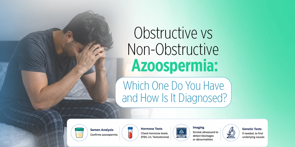
When a semen analysis returns showing zero sperm, the word “azoospermia” often dominates the conversation. But that word is only the beginning of the clinical picture — not the conclusion. The single most important question that follows is not whether a man has azoospermia, but which type.
Obstructive vs non-obstructive azoospermia are not simply two names for the same problem. They have different origins, different biological mechanisms, different genetic implications, different treatment pathways, and very different prognoses. Getting this distinction right is the essential first step in managing azoospermia — before any treatment decision is made.
This article focuses specifically on that distinction: what separates these two conditions at a biological level, how clinicians tell them apart, where diagnosis becomes genuinely difficult, and what the implications are for each man’s fertility journey.
The Fundamental Biological Difference
To understand obstructive vs non-obstructive azoospermia clearly, it helps to think of the male reproductive system as having two separate functions: production and delivery.
Obstructive Azoospermia (OA) is a delivery failure. The testes are producing sperm normally. The problem lies somewhere along the transport route — the epididymis, vas deferens, or ejaculatory ducts — where a physical blockage prevents that sperm from reaching the ejaculate. The factory is running; the road is blocked.
Non-Obstructive Azoospermia (NOA) is a production failure. There is no blockage in the transport system. The absence of sperm in the ejaculate is because the testes are not producing sufficient sperm — or, in some cases, producing none at all. The road is clear; the factory is not functioning as it should.
This distinction is not merely academic. It determines everything that follows — from which tests are ordered, to which treatments are viable, to what success rates a couple can realistically expect.
How Common Is Each Type?
Azoospermia affects approximately 1% of all men and accounts for around 10–15% of men evaluated for infertility. Within that population:
- Obstructive azoospermia accounts for approximately 40% of azoospermia cases
- Non-obstructive azoospermia accounts for approximately 60% of cases
These figures are consistent across multiple large studies and clinical reviews. NOA is therefore the more common type — and also the more clinically complex one.
Causes: What Drives Each Condition
Causes of Obstructive Azoospermia
The blockage in Obstructive azoospermia can arise from several sources:
- Vasectomy — the most common cause globally; a deliberate surgical block of the vas deferens
- Congenital bilateral absence of the vas deferens (CBAVD) — absence of the vas deferens from birth, strongly linked to CFTR gene mutations (the same gene associated with cystic fibrosis)
- Epididymal obstruction — from past infection (chlamydia, gonorrhoea, epididymitis), causing scarring that narrows or blocks the epididymal tubules
- Ejaculatory duct obstruction — from cysts, calcifications, or inflammation at the point where the ducts enter the urethra
- Iatrogenic injury — unintended damage to the vas deferens or epididymis during hernia repair, scrotal surgery, or other pelvic procedures
- Young’s Syndrome — a rare condition combining chronic respiratory infections with epididymal obstruction
Causes of Non-Obstructive Azoospermia
Non-obstructive azoospermia has a broader and often more complex set of underlying causes:
- Genetic conditions — including Klinefelter syndrome (47,XXY karyotype), Y-chromosome microdeletions (particularly in the AZF regions), and other chromosomal abnormalities; collectively, genetic factors account for a substantial proportion of NOA cases
- Cryptorchidism — undescended testes in childhood, which can permanently impair spermatogenesis even after surgical correction
- Varicocele — abnormally enlarged veins in the scrotum that raise testicular temperature and impair sperm production; a treatable but frequently overlooked cause
- Hypogonadotropic hypogonadism — a hormonal signalling failure where the pituitary gland does not produce adequate LH and FSH to stimulate the testes; notably, this is one of the few NOA subtypes that responds to medical treatment
- Chemotherapy or radiotherapy — gonadotoxic treatments that can permanently or temporarily damage testicular tissue
- Mumps orchitis — viral infection of the testes, which in post-pubertal males can cause significant and lasting damage to sperm-producing cells
- Idiopathic — in a meaningful proportion of NOA cases, no identifiable cause is found even after complete workup
The Diagnostic Process: How Clinicians Tell Them Apart
Distinguishing between the two types requires a structured, stepwise approach. No single test definitively separates OA from NOA in every case — diagnosis is built from a combination of findings.
Step 1: Physical Examination
The physical examination provides important early clues. An experienced clinician will assess:
- Testicular size and consistency — Normal-volume, firm testes suggest OA (production is intact). Small or soft testes are strongly suggestive of NOA (impaired spermatogenesis often causes testicular atrophy).
- Epididymis — Fullness or induration of the epididymis can indicate downstream obstruction consistent with OA.
- Vas deferens — The vas deferens should be palpable in the scrotum. Absence of the vas deferens on both sides is immediately diagnostic of CBAVD (an obstructive cause).
- Varicocele — A palpable or visible varicocele on examination raises the possibility of NOA due to impaired production.
Step 2: Semen Analysis and Semen Parameters
Beyond the sperm count itself, semen analysis provides useful diagnostic clues:
- Semen volume — Low semen volume (below 1.5 ml) alongside azoospermia can suggest ejaculatory duct obstruction or absence of seminal vesicles, pointing toward OA
- Semen pH — Acidic semen (below pH 7.0) in combination with low volume is a recognised marker for ejaculatory duct obstruction
- Fructose — Absence of fructose in the semen indicates that the seminal vesicles are not contributing to the ejaculate, a finding associated with CBAVD or ejaculatory duct obstruction
- Alpha-glucosidase — A marker of epididymal function; low levels can suggest epididymal obstruction
A retrospective study of 707 azoospermia patients identified semen volume, semen pH, and seminal plasma neutral alpha-glucosidase activity as three of the five most diagnostically useful parameters for distinguishing OA from NOA — without biopsy.
Step 3: Hormonal Profile
Blood hormone levels are among the most informative non-invasive tests for differentiating OA from NOA:
FSH (Follicle-Stimulating Hormone) FSH is produced by the pituitary gland and stimulates sperm production in the testes. When the testes are failing to produce sperm (NOA), the pituitary compensates by secreting more FSH — resulting in elevated levels. In obstructive azoospermia, because spermatogenesis is normal, FSH levels are typically within the normal range.
An elevated FSH in an azoospermic man is a strong indicator of NOA. A normal FSH does not confirm OA, but is consistent with it.
Inhibin B Inhibin B is secreted by the Sertoli cells of the testes — the cells that nurse developing sperm. Low inhibin B is a sensitive marker of impaired spermatogenesis and is frequently reduced in NOA, often more reliably than FSH alone.
Testosterone and LH Low testosterone combined with low LH points specifically toward hypogonadotropic hypogonadism — the hormonal form of NOA that is potentially treatable with gonadotropin therapy, rather than requiring sperm retrieval.
Step 4: Genetic Testing
Genetic testing is particularly important in NOA and in specific OA presentations:
Karyotype analysis (chromosomal testing) Recommended for all men with NOA. Klinefelter syndrome (47,XXY) is the most common chromosomal cause of NOA, affecting roughly 1 in 660 men. Men with Klinefelter syndrome may still have small foci of sperm production in the testes that can be retrieved with Micro-TESE.
Y-chromosome microdeletion analysis Deletions in specific regions of the Y chromosome (AZFa, AZFb, AZFc) impair or eliminate spermatogenesis. AZFa and AZFb deletions are associated with complete absence of sperm even on biopsy, making sperm retrieval unlikely to succeed. AZFc deletions leave open the possibility of retrieval. Knowing the deletion subtype before planning treatment prevents futile and unnecessary procedures.
CFTR gene mutation testing Recommended when CBAVD is identified (an obstructive cause). Men with CBAVD carry CFTR mutations, which can be passed to children conceived via IVF/ICSI. Genetic counselling before proceeding is essential in these cases.
Step 5: Scrotal and Transrectal Ultrasound
Imaging helps localise structural abnormalities:
- Scrotal ultrasound — assesses testicular size and echogenicity (texture), epididymal dilation, and varicocele
- Transrectal ultrasound (TRUS) — examines the prostate, ejaculatory ducts, and seminal vesicles for cysts or dilation suggesting ejaculatory duct obstruction
Step 6: Testicular Biopsy — The Definitive Test When Others Are Inconclusive
When clinical assessment, hormones, and imaging do not clearly distinguish OA from NOA, a testicular biopsy provides the definitive answer. Histological examination of testicular tissue shows:
- In OA — normal spermatogenesis with mature spermatozoa present in the seminiferous tubules, confirming that sperm production is intact
- In NOA — various patterns of impaired spermatogenesis: maturation arrest (sperm development halts at an early stage), Sertoli cell-only syndrome (no germ cells present at all), or hypospermatogenesis (reduced but present sperm production)
Critically, biopsy for diagnostic purposes should ideally be combined with sperm cryopreservation — if sperm are found, they are frozen immediately for potential future use in IVF/ICSI, avoiding a second surgical procedure.
The Diagnostic Grey Zone: When It Isn’t Clear-Cut
One aspect rarely discussed in patient-facing content is that the OA vs NOA distinction is not always straightforward. A proportion of men — particularly those with normal or borderline FSH, normal testicular size, and no identified obstruction — fall into a diagnostically ambiguous zone.
Recent research is exploring advanced tools to navigate this. A 2025 study evaluated a machine-learning model using combined clinical, hormonal, ultrasonographic, and semen data to classify azoospermia type with higher accuracy than individual parameters alone. While machine-learning diagnostic tools are not yet standard clinical practice, this research underscores that no single marker is infallible — and that experienced clinical judgement, integrating multiple data points, remains essential.
For patients in this grey zone, the biopsy remains the gold standard — but the timing and approach should be planned carefully, in coordination with the IVF/embryology team, to maximise the value of the procedure.
Side-by-Side Comparison: OA vs NOA at a Glance
| Feature | Obstructive Azoospermia (OA) | Non-Obstructive Azoospermia (NOA) |
|---|---|---|
| Core problem | Delivery blockage | Production failure |
| Sperm production | Normal | Impaired or absent |
| Testicular size | Usually normal | Often small/soft |
| FSH level | Usually normal | Typically elevated |
| Inhibin B | Usually normal | Often low |
| Common causes | Vasectomy, CBAVD, infection, surgery | Klinefelter, Y-deletion, cryptorchidism, varicocele |
| Genetic testing priority | CFTR (if CBAVD) | Karyotype + Y-microdeletion |
| Treatment options | Surgery or sperm retrieval + IVF/ICSI | Sperm retrieval (Micro-TESE) + IVF/ICSI; hormonal treatment in specific cases |
| Sperm retrieval success | High (production is intact) | Variable (10–60% depending on cause) |
| Risk of passing condition to offspring | CFTR mutations (if CBAVD) | Chromosomal/Y-deletion risk; genetic counselling essential |
Prognosis: How Do Outcomes Differ?
This is where the obstructive vs non-obstructive azoospermia distinction has the most direct impact on a couple’s planning.
For OA, the prognosis is generally favourable. Because sperm production is intact, sperm retrieval procedures yield sperm in the vast majority of cases. Combined with IVF/ICSI, clinical pregnancy rates per cycle are typically in the range of 40–60%. Surgical reconstruction in suitable candidates can restore natural sperm flow, potentially allowing conception without assisted reproduction.
For NOA, outcomes are more variable and depend heavily on the underlying cause. In men with hypospermatogenesis or AZFc deletions, Micro-TESE may still find usable sperm. In men with complete Sertoli cell-only syndrome or AZFa/AZFb deletions, retrieval is unlikely to succeed. A coordinated diagnostic approach — integrating reproductive urology, genetics, pathology, and embryology — is essential for setting realistic expectations, as outlined in published differential diagnosis guidelines for azoospermia (PMC / Int. J. Molecular Sciences). Across the full spectrum of NOA, sperm retrieval rates with Micro-TESE are reported in the range of 40–60% — but this varies considerably by cause, meaning pre-treatment genetic and histological data are critical.
Importantly, men with NOA due to hypogonadotropic hypogonadism represent a genuinely treatable subgroup — gonadotropin injections can stimulate the testes to resume sperm production in a meaningful proportion of these men, sometimes allowing sperm to appear in the ejaculate without any surgical retrieval.
Implications for Genetic Counselling
Both OA and NOA can carry genetic implications for children conceived through assisted reproduction — a topic that deserves more attention than it often receives.
In CBAVD-related OA, the CFTR mutations carried by the father can be passed to children. If the female partner is also a CFTR carrier (which occurs in approximately 1 in 25 people of European ancestry), there is a meaningful risk of the child developing cystic fibrosis. Partner carrier testing before IVF/ICSI is strongly recommended.
In NOA with chromosomal or Y-deletion causes, these genetic abnormalities can be transmitted to sons conceived via ICSI. Y-chromosome microdeletions are passed directly from father to son; Klinefelter syndrome (an extra X chromosome) occurs de novo rather than being inherited directly but requires careful counselling. Pre-implantation genetic testing (PGT) of embryos is an option some couples choose in this context.
At Best Life Fertility Center, genetic counselling is integrated into the diagnostic pathway — not an optional add-on.
Conclusion
The comparison of obstructive vs non-obstructive azoospermia is not a trivial clinical formality — it is the gateway to everything that follows. The type of azoospermia a man has determines which tests come next, which treatments are realistic, what genetic risks need to be considered, and what success rates a couple can honestly expect. Confusing the two, or proceeding to treatment without confirming the distinction, risks wasted time, unnecessary procedures, and avoidable disappointment.
At Best Life Fertility Center in Dubai, the differential diagnosis between obstructive and non-obstructive azoospermia is treated with the clinical rigour it deserves — a thorough, coordinated workup involving andrology, reproductive endocrinology, embryology, and genetic counselling, all in one place. If you or your partner have received a finding of azoospermia, the next step is not panic — it is precision.
Book your male fertility evaluation at Best Life Fertility Center →
Frequently Asked Questions
Q: Can you tell from a semen analysis alone whether azoospermia is obstructive or non-obstructive?
No. Semen analysis confirms zero sperm but cannot determine the cause; further hormonal, genetic, and imaging tests — sometimes biopsy — are needed.
Q: Is non-obstructive azoospermia always untreatable?
No. Men with hormonal causes (hypogonadotropic hypogonadism) can respond to medication. Others may have sperm retrieved via Micro-TESE for IVF/ICSI.
Q: Does FSH level alone confirm which type of azoospermia a man has?
Not definitively. Elevated FSH strongly suggests NOA, but some NOA men have normal FSH, making biopsy the gold standard in ambiguous cases.
Q: Are sons conceived via ICSI at risk of inheriting azoospermia?
Potentially yes, depending on the cause. Men with Y-chromosome deletions or CBAVD mutations should receive genetic counselling before proceeding with IVF/ICSI.
Q: How long does it take to get a definitive diagnosis of OA vs NOA in Dubai?
A comprehensive workup — semen analysis, hormones, imaging, and genetics — typically takes two to four weeks; biopsy results add one to two weeks.